Why Small Pets Stop Eating: Understanding Gastrointestinal Stasis in Rabbits and Rodents

Small pets like rabbits, guinea pigs and other rodents rely on a digestive system that never truly stops working. Their gut is designed to stay in constant motion, powered by a steady intake of fibre-rich foods throughout the day. When they suddenly refuse their favourite hay or stop nibbling altogether, it is far more than a simple change in appetite. In Singapore, gastrointestinal (GI) stasis is one of the most common emergency presentations for small pets. It is often the earliest, and sometimes, the only sign that something is seriously wrong.
Because these animals are prey species, they instinctively hide discomfort. A rabbit that seems only “a bit quiet” or a guinea pig that leaves its food untouched may already be experiencing gastrointestinal stasis, a condition where the gut slows down or comes to a halt. This may happen silently at first, but it can become dangerous in a matter of hours. Gas builds up, harmful bacteria multiply and the pet quickly becomes weak or dehydrated.
Understandably, it can be frightening to see your usually active companion turn away from food. The good news is that recognising this early warning sign can make all the difference. Quick action and timely veterinary attention often lead to a full recovery. Understanding why appetite loss is so urgent is the first step toward protecting your small pet’s health, and ensuring they stay bright, active and happily munching the way they should.
What is gastrointestinal stasis? — How the digestive slowdown develops
Gastrointestinal stasis, often called GI stasis, is a condition where the normal movement of food through the digestive system slows down or stops altogether [1]. For rabbits and many small rodents, this is especially dangerous because their gut depends on continuous motion to stay healthy. Unlike carnivores or even humans, their digestive tract is designed for a steady flow of fibrous food that keeps everything moving smoothly from stomach to colon.
In a healthy rabbit or rodent, gut motility works like a conveyor belt. As they graze throughout the day, mainly on hay, fibre fuels the natural contractions of the intestines. These contractions push food along, maintain proper bacterial balance and produce consistent droppings. This constant movement is essential; even short breaks in eating can interrupt the delicate system.
When gut motility slows, food begins to sit still in the stomach or intestines [2]. This leads to fermentation and gas buildup, which causes discomfort and makes the animal even less willing to eat. Over time, harmful bacteria may overgrow, dehydration worsens and the digestive slowdown becomes harder to reverse. What began as a small drop in appetite can quickly turn into a cycle where pain and reduced intake reinforce each other.
A major challenge in recognising GI stasis early is that rabbits and rodents are prey animals. They are hardwired to mask signs of weakness to avoid attracting predators. Instead of obvious distress, they may appear slightly quieter, less active or subtly change their eating habits. By the time an owner notices something is wrong, the condition may already be well underway. Understanding how silently GI stasis develops is key to catching it early and seeking the right care.
What causes gastrointestinal stasis in small pets
Gastrointestinal stasis is a symptom and not a diagnosis. When these small pets are unwell due to various reasons, gastrointestinal stasis develops.
The following are the more common causes that contribute to this condition:
- Low-fibre diet and inappropriate feeding — Too many pellets, treats or low-fibre foods reduce gut motility and disturb healthy bacterial balance.
- Dehydration — Insufficient water intake dries out intestinal contents, making them harder to pass.
- Pain from dental disease — Overgrown teeth, sharp spurs or jaw discomfort make eating painful, leading to reduced food intake.
- Stress and environmental change — Travel, loud noises, new pets or routine disruption can suppress appetite.
- Liver torsion, kidney failure — lead to pain, loss of appetite and dehydration
- Hair ingestion during moulting — Excess grooming increases hair intake, which can accumulate in the gut when fibre intake drops.
- Lack of exercise — Limited movement slows natural intestinal contractions.
- Post-surgical effects — Anaesthesia, pain or reduced mobility after a procedure can temporarily affect appetite.
- Temperature extremes — Heat stress or sudden temperature changes can slow activity and eating.
These factors often overlap and early detection plays an important role in preventing the condition from escalating.
GI stasis symptoms owners should not ignore
Gastrointestinal stasis often begins subtly and because rabbits and rodents instinctively mask discomfort, early signs can be easy to overlook [4]. Paying close attention to small changes in behaviour or routine helps you catch the condition before it becomes severe. Key symptoms include:
- Reduced appetite or refusal to eat — One of the earliest and most important warning signs.
- Small, dry or absent droppings — Indicates slowed or halted gut movement.
- Lethargy — A noticeable drop in activity or interest in normal routines.
- Hunched posture or grinding teeth — Common signs of abdominal pain or discomfort.
- Bloating or a firm abdomen — Gas buildup may cause visible swelling or tenderness.
- Hiding behaviour — Many small pets withdraw when they feel unwell.
- Sudden changes in drinking habits — Drinking significantly more or less than usual may signal an underlying issue.
Recognising these symptoms early gives your pet the best chance of a quick and full recovery.
Why GI stasis is dangerous — Complications of delayed care
Gastrointestinal stasis can progress quickly, which is why even a short delay in treatment can put a rabbit or rodent at serious risk. Once the gut slows down, a chain reaction begins inside the body that becomes harder to reverse with time. The following complications highlight why appetite loss should always be treated as an emergency:
- Dehydration and electrolyte imbalance — Reduced eating and drinking lead to rapid fluid loss, making gut movement even slower [5].
- Accumulation of gas in the stomach — Gas builds up as food ferments, causing painful bloating and additional pressure on internal organs.
- Overgrowth of harmful bacteria — Stagnant food allows bad bacteria to multiply, releasing toxins that worsen the condition.
- Liver fat mobilisation (hepatic lipidosis) — When small pets stop eating, the body breaks down fat rapidly, overwhelming the liver [6].
- Risk of death within hours to days — Without treatment, the cycle of pain, dehydration and bacterial imbalance can quickly become fatal.
Prompt veterinary care is essential because these complications escalate silently but rapidly.
How veterinarians diagnose gastrointestinal stasis
At Dr. Paws Vet Care, we take a thorough and gentle approach to diagnosing gastrointestinal stasis because early, accurate detection makes a significant difference in recovery. Our team uses a combination of physical assessment and diagnostic tools to confirm the condition and uncover the underlying reasons behind your pet’s appetite loss. This ensures that treatment addresses both the digestive slowdown and any contributing health issues. Key steps in our evaluation include:
- Thorough clinical history from owner
- Clinical examination — Assessing behaviour, posture and general appearance for signs of pain or distress.
- Abdominal palpation — Feeling the stomach and intestines for gas, firmness or abnormal masses.
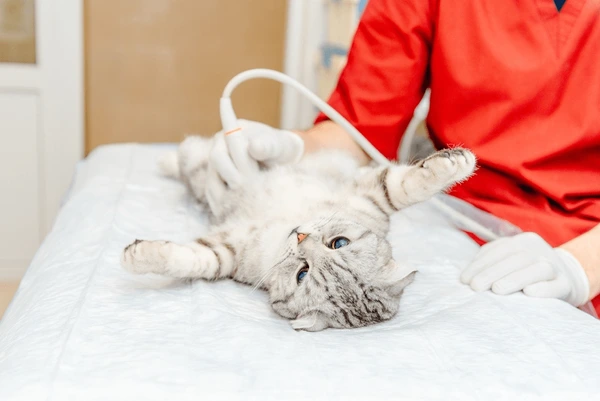
- X-rays or ultrasound — Imaging helps visualise gas patterns, stomach size, food accumulation or blockages. Liver torsion can be detected via ultrasound, although sometimes a referral for a CT scan may be required.
- Checking hydration status — Ensuring the pet is not dehydrated, which can worsen the condition.
- Dental evaluation — Identifying sharp spurs, overgrown teeth or jaw issues that may reduce appetite.
- Blood tests when needed — Used to assess organ function, detect infection or rule out metabolic causes.

Treatment options for gastrointestinal stasis
Treating gastrointestinal stasis requires prompt, targeted care and most pets respond well once the underlying issues are addressed. At Dr. Paws Vet Care, our goal is to stabilise your pet, relieve pain and safely restore normal gut movement. Treatment is always tailored to the severity of the condition and the animal’s overall health. Common veterinary interventions include:
- Fluid therapy (oral, subcutaneous, or IV) — Helps correct dehydration and soften intestinal contents, making them easier to move [7].
- Pain relief and anti-gas medications — Reduces discomfort and eases the pressure caused by gas buildup.
- Prokinetic drugs — Medications that gently stimulate the stomach and intestines to resume their normal contractions.
- Assisted feeding with critical-care formulas — Provides essential nutrition and encourages the digestive tract to start working again.
- Treatment of dental or underlying causes — Addressing tooth issues, infections or other medical conditions that triggered the slowdown.
- Warming, stress reduction and supportive care — Keeping the pet warm, hydrated and calm to support recovery.
Prompt treatment prevents complications and greatly improves the chances of a full recovery.
What you can do at home — Safe steps while waiting for vet care
GI stasis is an emergency which needs urgent attention.
While GI stasis always requires professional treatment, there are a few safe steps you can take at home to support your pet before reaching the clinic. These actions help keep your pet comfortable without risking further harm.
- Remove stressful triggers — Reduce noise, limit handling and create a quiet space.
- Keep your pet warm and calm — A cosy environment helps reduce stress-related gut slowdown.
- Offer favourite high-fibre foods — Encourage nibbling with fresh hay or leafy greens
- Avoid forcing water or medications — Syringe-feeding water or giving non-prescribed remedies can worsen the condition.
- Avoid over-the-counter treatments — Human or general pet medications are unsafe and may delay proper care.
- Call a vet urgently if symptoms persist — Early intervention is critical, especially if there is no eating, reduced droppings, or visible discomfort.
These steps are supportive only and professional treatment remains essential for reversing gastrointestinal stasis safely.
Prevention from gastrointestinal stasis — How to keep your rabbit or rodent’s gut healthy
Maintaining a healthy digestive system is one of the most effective ways to prevent gastrointestinal stasis. Simple, consistent care can keep your small pet’s gut moving well and reduce the risk of sudden appetite loss. Key preventive measures include:
- Provide unlimited access to high-quality hay every day.
- Feed species-appropriate, fibre-rich pellets in controlled portions.
- Ensure your pet always has clean, fresh water available.
- Encourage daily exercise and offer mental enrichment activities.
- Groom your pet regularly, especially during moulting periods.
- Schedule routine dental checks to catch early signs of tooth problems.
- Avoid making sudden changes to diet, housing, or environment.
- Maintain a calm, predictable routine to minimise stress.
When to see a vet for gastrointestinal stasis — Red flags that require emergency care
Certain symptoms indicate that gastrointestinal stasis may already be developing, and timely veterinary care becomes critical. If you notice any of the following warning signs, seek help immediately:
- Your pet has not eaten anything for 6–8 hours.
- There are no droppings present or droppings have suddenly stopped.
- The stomach appears visibly bloated or firm to the touch.
- Your pet is severely lethargic or unusually inactive.
- You observe teeth grinding or other signs of pain.
- Your pet refuses to drink or shows a sharp drop in water intake.
- There are sudden, unexplained changes in normal behaviour.
What small pets Dr Paws Vet Care sees
At Dr Paws Vet Care, our team treats a wide range of small mammals that are prone to gastrointestinal stasis and other digestive issues. We provide gentle, species-appropriate care for:
- Rabbits (all breeds)
- Guinea pigs
- Hamsters (Syrian, dwarf and hybrid)
- Gerbils
- Chinchillas
- Rats and mice
- Hedgehogs
- Other pocket pets commonly kept in Singapore
Our veterinarian and clinical team are experienced in recognising subtle signs of illness in prey species and provide diagnostics, treatment and ongoing management tailored to the needs of each animal.
Summary
Gastrointestinal stasis is one of the most urgent health issues faced by rabbits and small rodents and even a brief pause in eating can be the first sign that their digestive system is struggling. Understanding how delicately their gut functions, why appetite loss happens and which early symptoms to look out for helps owners act quickly and confidently. With the right diet, consistent routines, regular dental care and close attention to behaviour, many cases of GI stasis can be prevented. But when the condition does develop, prompt veterinary treatment makes a tremendous difference, restoring comfort, easing pain and getting the gut moving again.
If you’re worried that your rabbit or small pet has stopped eating, or you have noticed changes in droppings, behaviour or activity, don’t wait. Schedule a consultation with Dr. Paws Vet Care for prompt assessment, gentle handling and the right treatment to help your pet recover safely.
FAQ
Why did my rabbit stop eating suddenly?
A sudden loss of appetite in rabbits is often an early sign of gastrointestinal stasis. Pain, dental issues, stress, dehydration, low-fibre diets, infections or environmental change can all disrupt normal gut motility. Because rabbits mask discomfort, reduced eating is usually the first noticeable symptom and should be treated as urgent.
Is gastrointestinal stasis an emergency?
Yes. Gastrointestinal stasis is considered a veterinary emergency because the digestive tract can slow down or stop within hours. Gas buildup, dehydration and bacterial imbalance can develop quickly, leading to pain and potentially life-threatening complications if not treated promptly.
How fast can GI stasis become serious?
GI stasis can progress rapidly, sometimes within 6–12 hours from the first sign of appetite loss. As the gut slows, gas accumulates and the rabbit becomes increasingly uncomfortable, making them even less willing to eat. Early intervention significantly improves recovery and helps prevent severe complications.
Can GI stasis resolve without veterinary care?
It is unlikely and unsafe to rely on self-resolution. While mild cases may show temporary improvement, most require professional treatment to restore gut movement, correct dehydration and address underlying causes. Delaying care increases the risk of recurrence, worsening symptoms or life-threatening complications.
References
- doctormmdev. (2025, March 5). What is gastrointestinal stasis in rabbits? Animal Emergency PLLC. https://ervets4pets.doctormmdev3.com/what-is-gastrointestinal-stasis-in-rabbits/
- The Rabbit The Rabbit digestive system. https://rabbitwelfare.co.uk/wp-content/uploads/2023/05/article-ROWinter10p7.pdf
- Oglesbee, B. L., & Lord, B. (2020). Gastrointestinal diseases of rabbits. Ferrets, Rabbits, and Rodents, 174–187. https://doi.org/10.1016/B978-0-323-48435-0.00014-9
- Beuoy, C. (2024, June 4). Rabbit gastrointestinal stasis syndrome. Veterinary Medicine at Illinois. https://vetmed.illinois.edu/2024/06/04/rabbit-gi-stasis/
- Karin Allenspach, D. med vet. (2015). World small animal veterinary association world congress proceedings, 2010. VIN.Com. http://www.vin.com/doc/?id=6697938
- Karin Allenspach, D. med vet. (2015). World small animal veterinary association world congress proceedings, 2010. VIN.Com. http://www.vin.com/doc/?id=6697938
- Managing gastrointestinal stasis in small mammals. (2022, December 9). https://improveinternational.com/uk/clinical-library/managing-gastrointestinal-stasis-in-small-mammals